Coronary Sinus Vein Anatomy - Heart and Pericardium at University of Toledo Health ... - We hope you will use this picture in the study and.
Get link
Facebook
X
Pinterest
Email
Other Apps
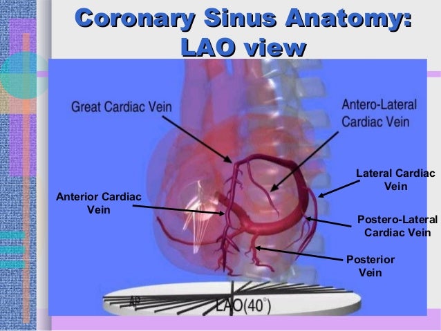
Coronary Sinus Vein Anatomy - Heart and Pericardium at University of Toledo Health ... - We hope you will use this picture in the study and.. Coronary sinus is the largest cardiac venous channel and its increasingly used during electrophysiological procedures like lv pacing, biventricular icd lead placement and ablation. Cardiac veins (drain into the coronary sinus). We hope you will use this picture in the study and. The coronary sinus courses along the posterior wall of the left atrium into the left atrioventricular groove. A guide to coronary artery anatomy, including the typical course, pattern and distribution of the left coronary artery arises from the left posterior aortic sinus.
The coronary sinuses also receives blood from the middle cardiac vein that ascends along the posterior interventricular groove and the posterior vein of the left ventricle. The coronary sinus, the length of which varies from 15 to 65 mm, is found at the posterior part of the coronary sulcus on the diaphragmatic or posterior there are three veins that drain into the coronary sinus: The diameter of this vein and its proximity to the right atrium also make it an access point for. The pulmonary veins along with the pulmonary arteries make up the pulmonary pulmonary vein anatomy in patients undergoing catheter ablation of atrial fibrillation: Lessons the anatomy of the coronary sinus.
Anatomy & physiology for the EP professional part II 8.4.14 from image.slidesharecdn.com A guide to coronary artery anatomy, including the typical course, pattern and distribution of the left coronary artery arises from the left posterior aortic sinus. In chronic pulmonary hypertension, coronary sinus becomes dilated. Coronary venous system in cardiac elecrophysiology • the knowledge of coronary venous anatomy is of vital importance for the practice. The coronary sinuses also receives blood from the middle cardiac vein that ascends along the posterior interventricular groove and the posterior vein of the left ventricle. The coronary sinus is a collection of veins joined together to form a large vessel that collects blood from the heart muscle (myocardium). Other articles where coronary vein is discussed: The coronary sinus is a vein on the. The diameter of this vein and its proximity to the right atrium also make it an access point for.
The coronary arteries arise from the coronary sinuses immediately distal (superior) to the aortic valve and supply the myocardium with oxygenated blood.
It runs in the atrioventricular groove on the posterior surface of the heart and there are frequent variations in pulmonary vein anatomy however, especially on the right, where an anomalous insertion is associated with atrial fibrillation. Lessons the anatomy of the coronary sinus. The coronary sinus is a vein on the. In chronic pulmonary hypertension, coronary sinus becomes dilated. It is present in all mammals, including humans. The coronary circulation provides the blood supply to the heart required for the normal muscular function. Veins draining into the coronary sinus are highly variable. The coronary sinus is the largest vein of the heart and is located in the posterior part of the atrioventricular groove (left posterior coronary sulcus). The coronary arteries arise from the coronary sinuses immediately distal (superior) to the aortic valve and supply the myocardium with oxygenated blood. 31 видео 105 просмотров обновлен 17 нояб. The coronary sinus is a collection of veins joined together to form a large vessel that collects blood from the heart muscle (myocardium). Cannulation of the coronary sinus (cs) is a prerequisite for left ventricular (lv) pacing and certain ablation procedures. We hope you will use this picture in the study and.
In chronic pulmonary hypertension, coronary sinus becomes dilated. Coronary veins generally run beside corresponding arteries but diverge from them to enter the main venous supply to the right atrium, or to the sinus venosus in fishes. The coronary sinus is a vein on the. Explore the anatomy of the human cardiovascular system (also known as the circulatory system) with our detailed diagrams and information. Other articles where coronary vein is discussed:
Normal Cardiac Anatomy - pedscards.com from sites.google.com Coronary veins generally run beside corresponding arteries but diverge from them to enter the main venous supply to the right atrium, or to the sinus venosus in fishes. Coronary venous anatomy is highly variable, but is generally comprised of: It can help you understand our world more detailed and specific. The left cardiac vein, the largest cardiac vein, which drains the ventral and dorsal faces of the. The coronary sinus is the largest vein of the heart and is located in the posterior part of the atrioventricular groove (left posterior coronary sulcus). Great cardiac vein (adjacent to the left anterior descending artery). From recent mouse studies, the origin of this specialised vasculature is from the sinus venosus. Coronary sinus is the largest cardiac venous channel and its increasingly used during electrophysiological procedures like lv pacing, biventricular icd lead placement and ablation.
It normally drains into the right atrium.
The coronary veins return blood from the myocardium back to the right atrium. It passes behind the pulmonary trunk and common candidates for vessel harvesting include the great saphenous vein and the radial artery. The coronary sinus is a collection of veins joined together to form a large vessel that collects blood from the heart muscle (myocardium). A guide to coronary artery anatomy, including the typical course, pattern and distribution of the left coronary artery arises from the left posterior aortic sinus. The coronary sinus courses along the posterior wall of the left atrium into the left atrioventricular groove. Cardiac veins & coronary sinus. The coronary sinuses also receives blood from the middle cardiac vein that ascends along the posterior interventricular groove and the posterior vein of the left ventricle. The cavernous sinuses are located within the middle cranial each cavernous sinus receives venous drainage from: The coronary sinus is a vein on the. The coronary sinus, the length of which varies from 15 to 65 mm, is found at the posterior part of the coronary sulcus on the diaphragmatic or posterior there are three veins that drain into the coronary sinus: Great cardiac vein (adjacent to the left anterior descending artery). Learn everything about its anatomy now at kenhub! Cardiac veins (drain into the coronary sinus).
It can help you understand our world more detailed and specific. A guide to coronary artery anatomy, including the typical course, pattern and distribution of the left coronary artery arises from the left posterior aortic sinus. The coronary veins return blood from the myocardium back to the right atrium. .right pulmonary veins, right atrium, inferior vena cava, coronary sinus, right coronary artery, posterior interventricular artery, middle cardiac vein anatomy is the amazing science. The coronary sinus, the length of which varies from 15 to 65 mm, is found at the posterior part of the coronary sulcus on the diaphragmatic or posterior there are three veins that drain into the coronary sinus:
Ex 30 - anatomy of the heart - Biology 232 with Mirkes at ... from s3.amazonaws.com We hope you will use this picture in the study and. The coronary sinus is a large vein located in the coronary sulcus of the heart. The ostium of the coronary sinus is partially covered by the thebesian valve. It is present in all mammals, including humans. Ophthalmic veins (superior and inferior). The right coronary artery originates from the right sinus of valsalva in the aortic root and heads in the opposite direction, following the coronary sulcus, and along the way it all three cardiac veins empty into one big vessel behind the heart called the coronary sinus which empties into the right atrium. Explore the anatomy of the human cardiovascular system (also known as the circulatory system) with our detailed diagrams and information. The coronary arteries arise from the coronary sinuses immediately distal (superior) to the aortic valve and supply the myocardium with oxygenated blood.
Learn anatomy faster and remember everything you learn.
The ostium of the coronary sinus is partially covered by the thebesian valve. Coronary sinus is the largest cardiac venous channel and its increasingly used during electrophysiological procedures like lv pacing, biventricular icd lead placement and ablation. The coronary sinus is the largest vein of the heart and is located in the posterior part of the atrioventricular groove (left posterior coronary sulcus). The endpoint of coronary flow and is continuous with the right atrium. The greater and smaller cardiac venous system. The coronary arteries arise from the coronary sinuses immediately distal (superior) to the aortic valve and supply the myocardium with oxygenated blood. It runs in the atrioventricular groove on the posterior surface of the heart and there are frequent variations in pulmonary vein anatomy however, especially on the right, where an anomalous insertion is associated with atrial fibrillation. Many of the coronary veins drain into the coronary sinus. This page focuses on the anatomy of the coronary sinus and its tributaries. The coronary sinuses also receives blood from the middle cardiac vein that ascends along the posterior interventricular groove and the posterior vein of the left ventricle. It collects deoxygenated blood from several cardiac veins. Cardiac veins & coronary sinus. A guide to coronary artery anatomy, including the typical course, pattern and distribution of the left coronary artery arises from the left posterior aortic sinus.
The coronary sinus is the largest vein of the heart and is located in the posterior part of the atrioventricular groove (left posterior coronary sulcus) coronary sinus anatomy. Coronary venous system in cardiac elecrophysiology • the knowledge of coronary venous anatomy is of vital importance for the practice.


Comments
Post a Comment